

How Does Our Teeth Enamel Repair
| Molar enamel | |
|---|---|
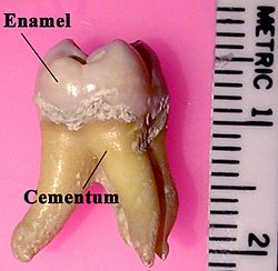 Labeled molar | |
| Details | |
| Identifiers | |
| Latin | enamelum |
| MeSH | D003743 |
| TA98 | A05.1.03.056 |
| TA2 | 938 |
| FMA | 55629 |
| Anatomical terminology [edit on Wikidata] | |
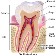
Parts of a tooth, including the enamel (cross section).
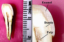
Tooth enamel is ane of the four major tissues that make up the tooth in humans and many other animals, including some species of fish. Information technology makes up the normally visible office of the tooth, covering the crown. The other major tissues are dentin, cementum, and dental pulp. It is a very hard, white to off-white, highly mineralised substance that acts as a barrier to protect the tooth but can become susceptible to degradation, especially by acids from food and drink. Calcium hardens the tooth enamel. In rare circumstances enamel fails to form, leaving the underlying dentin exposed on the surface.[1]
Features [edit]
Enamel is the hardest substance in the human trunk and contains the highest per centum of minerals (at 96%),[ii] with water and organic material composing the rest.[3] The primary mineral is hydroxyapatite, which is a crystalline calcium phosphate.[four] Enamel is formed on the tooth while the tooth develops within the jaw bone before it erupts into the mouth. Once fully formed, enamel does not contain claret vessels or fretfulness, and is non fabricated of cells. Remineralisation of teeth tin can repair damage to the tooth to a sure degree but damage across that cannot be repaired past the body. The maintenance and repair of human being tooth enamel is i of the primary concerns of dentistry.
In humans, enamel varies in thickness over the surface of the molar, often thickest at the cusp, up to ii.5 mm, and thinnest at its border with the cementum at the cementoenamel junction (CEJ).[5]
The normal color of enamel varies from light yellow to grayish (bluish) white. At the edges of teeth where there is no dentin underlying the enamel, the colour sometimes has a slightly blue or translucent off-white tone, easily observable on the upper incisors. Since enamel is semitranslucent, the color of dentin and whatever material underneath the enamel strongly affects the appearance of a tooth. The enamel on primary teeth has a more than opaque crystalline grade and thus appears whiter than on permanent teeth.
The big corporeality of mineral in enamel accounts non only for its forcefulness but also for its brittleness.[6] Tooth enamel ranks 5 on Mohs hardness scale (between steel and titanium) and has a Young's modulus of 83 GPa.[4] Dentin, less mineralized and less brittle, 3–4 in hardness, compensates for enamel and is necessary as a support.[7] On radiographs, the differences in the mineralization of dissimilar portions of the tooth and surrounding periodontium can be noted; enamel appears lighter than dentin or pulp since it is denser than both and more radiopaque.[8]
Enamel does not contain collagen, as found in other hard tissues such as dentin and bone, just it does contain two unique classes of proteins: amelogenins and enamelins. While the role of these proteins is not fully understood, it is believed that they aid in the development of enamel by serving as a framework for minerals to form on, amongst other functions.[half-dozen] In one case it is mature, enamel is almost totally without the softer organic matter. Enamel is avascular and has no nerve supply within it and is not renewed, still, it is non a static tissue as it tin can undergo mineralization changes.[9]
Structure [edit]
The bones unit of measurement of enamel is called an enamel rod.[seven] Measuring 4–8 μm in diameter, an enamel rod, formally called an enamel prism, is a tightly packed mass of hydroxyapatite crystallites in an organized pattern.[two] In cross section, it is best compared to a keyhole, with the top, or caput, oriented toward the crown of the molar, and the bottom, or tail, oriented toward the root of the tooth.
The arrangement of the crystallites within each enamel rod is highly circuitous. Both ameloblasts (the cells which initiate enamel formation) and Tomes' processes affect the crystallites' blueprint. Enamel crystallites in the head of the enamel rod are oriented parallel to the long axis of the rod.[2] [5] When found in the tail of the enamel rod, the crystallites' orientation diverges slightly (65 degrees) from the long centrality.[2]
The organisation of enamel rods is understood more clearly than their internal structure. Enamel rods are found in rows forth the tooth, and inside each row, the long axis of the enamel rod is generally perpendicular to the underlying dentin.[x] In permanent teeth, the enamel rods most the cementoenamel junction (CEJ) tilt slightly toward the root of the tooth. Agreement enamel orientation is very important in restorative dentistry, because enamel unsupported past underlying dentin is decumbent to fracture.[x]
The area around the enamel rod is known as interrod enamel. Interrod enamel has the same composition as enamel rod, however a histologic distinction is fabricated between the two because crystallite orientation is different in each.[5] The border where the crystallites of enamel rods and crystallites of interrod enamel come across is chosen the rod sheath.[10]
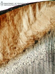
Striae of Retzius are incremental lines that appear brown in a stained section of mature enamel. These lines are composed of bands or cantankerous striations on the enamel rods that, when combined in longitudinal sections, seem to traverse the enamel rods.[ten] Formed from changes in bore of Tomes' processes, these incremental lines demonstrate the growth of enamel, similar to the annual rings on a tree on transverse sections of enamel. The exact mechanism that produces these lines is still being debated. Some researchers hypothesize that the lines are a result of the diurnal (circadian), or 24-60 minutes, metabolic rhythm of the ameloblasts producing the enamel matrix, which consists of an agile secretory work period followed by an inactive rest flow during tooth development. Thus, each band on the enamel rod demonstrates the work/remainder pattern of the ameloblasts that mostly occurs over a span of a calendar week.[xi]
Perikymata which are associated with the Striae are shallow grooves noted clinically on the nonmasticatory surfaces of some teeth in the oral cavity.[6] Perikymata are usually lost through tooth wear, except on the protected cervical regions of some teeth, particularly the permanent maxillary central incisors, canines, and first premolars, and may exist confused as dental calculus.[11] Darker than the other incremental lines, the neonatal line is an incremental line that separates enamel formed before and afterwards nascency.[12] The neonatal line marks the stress or trauma experienced by the ameloblasts during birth, once again illustrating the sensitivity of the ameloblasts every bit they course enamel matrix. Equally 1 would expect, the neonatal line is found in all primary teeth and in the larger cusps of the permanent beginning molars. They contain irregular structures of enamel prisms with matted crystallite arrangements basically formed by the abrupt bending of the prisms towards the root; normally, the prisms gradually bent back again to regain their previous orientation.[11]
Gnarled enamel is found at the cusps of teeth.[iii] Its twisted appearance results from the orientation of enamel rods and the rows in which they lie.
Evolution [edit]

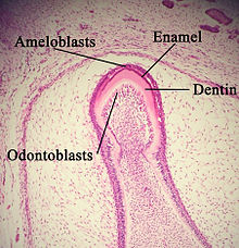
Histologic slide showing a developing tooth. The mouth would be in the surface area of infinite at the top of the movie.
Enamel formation is role of the overall process of molar development. Nether a microscope, different cellular aggregations are identifiable within the tissues of a developing tooth, including structures known as the enamel organ, dental lamina, and dental papilla.[13] The generally recognized stages of tooth development are the bud stage, cap stage, bong stage, and crown, or calcification, stage. Enamel formation is kickoff seen in the crown phase.
Amelogenesis, or enamel germination, occurs after the first establishment of dentin, via cells known as ameloblasts. Human enamel forms at a charge per unit of effectually 4 μm per day, offset at the futurity location of cusps, around the third or fourth month of pregnancy.[10] Every bit in all human processes, the cosmos of enamel is complex, but can more often than not be divided into two stages.[3] The beginning phase, called the secretory phase, involves proteins and an organic matrix forming a partially mineralized enamel. The second phase, called the maturation stage, completes enamel mineralization.
Histologic slide showing enamel germination
In the secretory phase, ameloblasts are polarized columnar cells. In the rough endoplasmic reticulum of these cells, enamel proteins are released into the surrounding area and contribute to what is known as the enamel matrix, which is and then partially mineralized past the enzyme alkaline phosphatase.[14] When this commencement layer is formed, the ameloblasts movement away from the dentin, allowing for the development of Tomes' processes at the upmost pole of the prison cell. Enamel germination continues effectually the bordering ameloblasts, resulting in a walled area, or pit, that houses a Tomes' process, and also around the end of each Tomes' process, resulting in a degradation of enamel matrix inside of each pit.[iii] The matrix within the pit will somewhen become an enamel rod, and the walls will somewhen become interrod enamel. The only distinguishing cistron between the two is the orientation of the calcium phosphate crystallites.
In the maturation stage, the ameloblasts send substances used in the formation of enamel. Histologically, the most notable attribute of this phase is that these cells become striated, or have a ruffled border.[14] These signs demonstrate that the ameloblasts have inverse their role from production, as in the secretory stage, to transportation. Proteins used for the terminal mineralization process compose most of the transported cloth. The noteworthy proteins involved are amelogenins, ameloblastins, enamelins, and tuftelins. How these proteins are secreted into the enamel structure is still unknown; other proteins, such as the Wnt signaling components BCL9 and Pygopus, have been implicated in this process.[xv] During this process, amelogenins and ameloblastins are removed afterward apply, leaving enamelins and tuftelin in the enamel.[sixteen] Past the end of this phase, the enamel has completed its mineralization.
At some indicate before the tooth erupts into the mouth, but after the maturation phase, the ameloblasts are cleaved downwardly. Consequently, enamel, unlike many other tissues of the torso, has no way to regenerate itself.[17] After destruction of enamel from decay or injury, neither the torso nor a dentist can restore the enamel tissue. Enamel can be afflicted farther by not-pathologic processes.
Enamel is covered by diverse structures in relation to the evolution of molar:
-
- Nasmyth membrane or enamel cuticle, construction of embryological origin is composed of keratin which gives ascension to the enamel organ.[18] [19]
- Acquired pellicle, structure acquired after tooth eruption is composed of food debris, calculus, dental plaque (organic picture show).[20]
Progress of enamel formation for chief teeth[21]
| Corporeality of enamel formed at nascence | Enamel mineralization completed | ||
|---|---|---|---|
| Primary maxillary tooth | Central incisor | 5/6 | ane.5 months subsequently nascence |
| Lateral incisor | 2/3 | 2.5 months later nascence | |
| Canine | one/3 | 9 months subsequently birth | |
| 1st tooth | Cusps united; occlusal completely calcified and 1/ii to 3/4 crown meridian | 6 months after nativity | |
| 2nd molar | Cusps united; occlusal incompletely calcified; calcified tissue covers 1/five to i⁄4 crown top | 11 months after nativity | |
| Chief mandibular tooth | Central incisor | 3/5 | 2.5 months later on nascence |
| Lateral incisor | 3/5 | 3 months after nascence | |
| Canine | 1/iii | 9 months after nascency | |
| 1st tooth | Cusps united; occlusal completely calcified | 5.five months after nascence | |
| 2nd molar | Cusps united; occlusal incompletely calcified | 10 months afterward birth | |
Enamel loss [edit]
The high mineral content of enamel, which makes this tissue the hardest in the human body, also makes it demineralize in a procedure that often occurs every bit dental caries, otherwise known as cavities.[13] Demineralization occurs for several reasons, simply the almost important cause of tooth disuse is the ingestion of fermentable carbohydrates.[ citation needed ] Tooth cavities are caused when acids dissolve molar enamel:[22] Enamel is as well lost through tooth wearable and enamel fractures.[23]
-
- Ca10(POiv)6(OH)2(s) + 8H+(aq) → 10Ca2+(aq) + 6HPO4 2−(aq) + 2H2O(fifty)
Sugars and acids from candies, soft drinks, and fruit juices play a significant role in tooth decay, and consequently in enamel destruction.[24] The mouth contains a bang-up number and variety of bacteria, and when sucrose, the most common of sugars, coats the surface of the oral fissure, some intraoral bacteria interact with it and form lactic acid, which decreases the pH in the mouth.[25] The disquisitional pH for tooth enamel is more often than not accepted to be pH 5.5. When acids are present and the critical pH is reached, the hydroxyapatite crystallites of enamel demineralize, allowing for greater bacterial invasion deeper into the tooth. The near important bacterium involved with tooth disuse is Streptococcus mutans, but the number and type of bacteria varies with the progress of molar destruction.[25]
Furthermore, tooth morphology dictates that the most common site for the initiation of dental caries is in the deep grooves, pits, and fissures of enamel.[ citation needed ] This is expected because these locations are impossible to reach with a toothbrush and allow for bacteria to reside there. When demineralization of enamel occurs, a dentist can use a sharp instrument, such every bit a dental explorer, and "feel a stick" at the location of the decay. As enamel continues to get less mineralized and is unable to preclude the inroad of bacteria, the underlying dentin becomes affected as well. When dentin, which commonly supports enamel, is destroyed past a physiologic condition or by disuse, enamel is unable to compensate for its brittleness and breaks abroad from the molar easily.

The furnishings of bruxism on an anterior tooth, revealing the dentin and lurid which are normally hidden by enamel
The extent to which tooth disuse is likely, known as cariogenicity, depends on factors such as how long the carbohydrate remains in the rima oris. Contrary to common belief, information technology is non the corporeality of sugar ingested but the frequency of carbohydrate ingestion that is the most important factor in the causation of tooth decay.[26] When the pH in the oral cavity initially decreases from the ingestion of sugars, the enamel is demineralized and left vulnerable for about 30 minutes. Eating a greater quantity of sugar in one sitting does not increment the fourth dimension of demineralization. Similarly, eating a bottom quantity of sugar in one sitting does not decrease the fourth dimension of demineralization. Thus, eating a great quantity of sugar at one time in the 24-hour interval is less detrimental than is a very small quantity ingested in many intervals throughout the day. For case, in terms of oral health, it is improve to eat a unmarried dessert at dinner time than to snack on a bag of candy throughout the day.
In addition to bacterial invasion, enamel is also susceptible to other destructive forces. Bruxism, likewise known as clenching of or grinding on teeth, destroys enamel very rapidly. The vesture rate of enamel, chosen attrition, is eight micrometers a twelvemonth from normal factors.[ citation needed ] A common misconception is that enamel wears away mostly from chewing, only actually teeth rarely bear on during chewing. Furthermore, normal tooth contact is compensated physiologically by the periodontal ligaments (pdl) and the organisation of dental occlusion. The truly destructive forces are the parafunctional movements, as plant in bruxism, which can cause irreversible impairment to the enamel.
Other nonbacterial processes of enamel destruction include abrasion (involving foreign elements, such as toothbrushes), erosion (involving chemic processes, such as dissolving by soft drinks[27] or lemon and other juices), and possibly abfraction (involving compressive and tensile forces).[ citation needed ]
Though enamel is described as tough, it has a similar brittleness to drinking glass, making information technology, different other natural crack-resistant laminate structures such as shell and nacre, vulnerable to fracture. In spite of this information technology can withstand bite forces every bit loftier equally ane,000 N many times a day during chewing.[28] [29] This resistance is due in part to the microstructure of enamel which contains enamel tufts that stabilize such fractures at the dentinoenamel junction.[30] The configuration of the tooth besides acts to reduce the tensile stresses that cause fractures during biting.[30]
Gastroesophageal reflux disease can besides lead to enamel loss, every bit acid refluxes up the esophagus and into the rima oris, occurring nearly during overnight sleep.
Oral hygiene [edit]
Considering enamel is vulnerable to demineralization, prevention of molar decay is the all-time way to maintain the health of teeth. Nearly countries have wide use of toothbrushes, which can reduce the number of dental biofilm and nutrient particles on enamel. In isolated societies that do non take access to toothbrushes, information technology is mutual for those people to utilize other objects, such as sticks, to clean their teeth. In betwixt 2 adjacent teeth, floss is used to wipe the enamel surfaces gratuitous of plaque and food particles to discourage bacterial growth. Although neither floss nor toothbrushes can penetrate the deep grooves and pits of enamel, proficient general oral-wellness habits can commonly forbid enough bacterial growth to keep tooth decay from starting. Structural integrity of the enamel is genetic, and so is its predisposition to demineralization or assail from bacteria.[xv]
Fluoride remineralization [edit]
Fluoride catalyzes the diffusion of calcium and phosphate into the tooth surface, which in turn remineralizes the crystalline structures in a dental cavity. The remineralized tooth surfaces contain fluoridated hydroxyapatite and fluorapatite, which resist acid set on much meliorate than the original molar did.[31] Fluoride therapy is used to assistance prevent dental decay.

Mutual dentistry trays filled with fluoride foam
Fluoride ions, as an antimicrobial, may activate bacterial genes associated with fluoride riboswitches.[32] [ unreliable medical source? ] The combination of fluoride ions and QAS (quaternary ammonium salts) was found to have a stronger antimicrobial effect on many oral leaner associated with dental decay, including S. mutans.
Fluoride in drinking water [edit]
Near dental professionals and organizations concord that the inclusion of fluoride in public water has been one of the near effective methods of decreasing the prevalence of molar decay.[33] Fluoride can be found in many locations naturally, such as the ocean and other water sources. The recommended dosage of fluoride in drinking water does not depend on air temperature.[34]
Some groups have spoken out against fluoridated drinking water, for reasons such as the neurotoxicity of fluoride or the damage fluoride can practice as fluorosis. Fluorosis is a condition resulting from the overexposure to fluoride, especially between the ages of 6 months and 5 years, and appears as mottled enamel.[3] Consequently, the teeth wait unsightly, although the incidence of dental decay in those teeth is very small-scale. Where fluoride is found naturally in high concentrations, filters are often used to subtract the amount of fluoride in water. For this reason, codes have been developed by dental professionals to limit the amount of fluoride a person should accept.[35] These codes are supported by the American Dental Association and the American Academy of Pediatric Dentistry;
Furthermore, whereas topical fluoride, found in toothpaste and mouthwashes, does not crusade fluorosis, its effects are now considered more important than those of systemic fluoride, such as when drinking fluorinated h2o.[36] Withal, systemic fluoride works topically equally well with fluoride levels in saliva increase also when drinking fluoridated h2o. Lately, dental professionals are looking for other ways to present fluoride (such as in varnish) or other mineralizing products such as Baggy calcium phosphate to the community in the class of topical procedures, either washed past professionals or self-administered. Mineralization of the incipient lesion instead of restoration later is a prime number goal of most dental professionals.
Regrowth [edit]
UK scientists at Bristol University and the Leeds Dental Institute take developed gels which tin can regenerate decayed or damaged molar enamel. A peptide hydrogel is applied to the molar. This forms into a protein scaffold onto which new enamel-forming calcium is deposited from the saliva. The scientists claim to have seen "highly significant" levels of repair in which signs of decay have been reversed months after a single application of the compound.[37] [38] [ unreliable medical source? ]
Researchers at the University of Southern California take adult an amelogenin-chitosan peptide hydrogel which entraps calcium and phosphorus mineral ions from saliva, forming a highly oriented enamel-like layer, restoring up to 80% of the hardness of normal enamel.[39] [40] [ unreliable medical source? ]
In 2019 Chinese scientists constitute that mixing calcium and phosphate ions with triethylamine in an alcohol solution causes enamel to grow with the same construction every bit teeth (a biomimetic remineralization).[41] [ unreliable medical source? ]
Dental procedures [edit]

An 10-ray showing enamel and dentin replaced by an amalgam restoration
Dental restorations [edit]
Most dental restorations involve the removal of enamel. Frequently, the purpose of removal is to gain access to the underlying disuse in the dentin or inflammation in the lurid. This is typically the example in amalgam restorations and endodontic treatment.
Nonetheless, enamel tin sometimes be removed earlier in that location is any decay present. The most popular case is the dental sealant. In the past, the process of placing dental sealants involved removing enamel in the deep fissures and grooves of a molar, followed by replacing it with a restorative fabric.[42] Presently, information technology is more than mutual to only remove rust-covered enamel if present. In spite of this, there are all the same cases where deep fissures and grooves in enamel are removed in order to prevent disuse, and a sealant may or may not be placed depending on the situation. Sealants are unique in that they are preventative restorations for protection from futurity disuse, and accept been shown to reduce the risk of decay past 55% over 7 years.[43]
Aesthetics is another reason for the removal of enamel. Removing enamel is necessary when placing crowns and veneers to enhance the advent of teeth. In both of these instances, when unsupported by underlying dentin, that portion of the enamel is more vulnerable to fracture.[44]
Acid-etching techniques [edit]
Invented in 1955, acid-etching employs dental etchants and is used frequently when bonding dental restoration to teeth.[45] This is important for long-term use of some materials, such equally composites and sealants.[xiii] By dissolving minerals in enamel, etchants remove the outer 10 micrometers on the enamel surface and make a porous layer five–50 micrometers deep.[46] This roughens the enamel microscopically and results in a greater surface surface area on which to bond.
The effects of acid-carving on enamel can vary. Important variables are the amount of time the etchant is applied, the type of etchant used, and the electric current status of the enamel.[46]
There are 3 types of patterns formed by acrid-etching.[46] Blazon 1 is a pattern where predominantly the enamel rods are dissolved; type 2 is a blueprint where predominantly the surface area around the enamel rods are dissolved; and type 3 is a design where there is no bear witness left of any enamel rods. As well concluding that type ane is the most favorable pattern and type 3 the least, the caption for these different patterns is not known for certain but is nearly commonly attributed to different crystallite orientation in the enamel.[three]
Tooth whitening [edit]
The discoloration of teeth over time tin upshot from exposure to substances such every bit tobacco, coffee, and tea.[47] The staining occurs in the interprismatic region internally on the enamel, which causes the molar to appear darker or more xanthous overall. In a perfect country, enamel is colorless, simply it does reflect underlying tooth structure with its stains since light reflection properties of the molar are low.
Tooth whitening or tooth bleaching procedures attempt to lighten a tooth's colour in either of two ways: by chemical or mechanical activity. Working chemically, a bleaching agent is used to carry out an oxidation reaction in the enamel and dentin.[48] The agents most unremarkably used to intrinsically change the color of teeth are hydrogen peroxide and carbamide peroxide. Oxygen radicals from the peroxide in the whitening agents contact the stains in the interprismatic spaces within the enamel layer. When this occurs, stains volition exist bleached and the teeth now appear lighter in color. Teeth not just appear whiter but also reverberate light in increased amounts, which makes the teeth appear brighter equally well. Studies prove that whitening does not produce any ultrastructural or microhardness changes in the dental tissues.[8]
Studies testify that patients who have whitened their teeth take better intendance of them.[49] Yet, a molar whitening product with an overall low pH can put enamel at hazard for decay or destruction past demineralization. Consequently, intendance should exist taken and risk evaluated when choosing a product which is very acidic.[50] Molar whiteners in toothpastes piece of work through a mechanical action. They have mild abrasives which aid in the removal of stains on enamel. Although this can be an effective method, information technology does not alter the intrinsic color of teeth. Microabrasion techniques employ both methods. An acid is used beginning to weaken the outer 22–27 micrometers of enamel in club to weaken it enough for the subsequent annoying force.[51] This allows for removal of superficial stains in the enamel. If the discoloration is deeper or in the dentin, this method of tooth whitening will non be successful.
[edit]
Irreversible enamel defects acquired by an untreated celiac disease. They may be the only inkling to its diagnosis, even in absence of gastrointestinal symptoms, but are often confused with fluorosis, tetracycline discoloration, or other causes.[52] [53] [54] The National Institutes of Health include a dental exam in the diagnostic protocol of celiac illness.[52]
There are 14 different types of amelogenesis imperfecta.[3] The hypocalcification type, which is the most common, is an autosomal ascendant condition that results in enamel that is not completely mineralized.[55] Consequently, enamel easily flakes off the teeth, which announced yellow considering of the revealed dentin. The hypoplastic blazon is X-linked and results in normal enamel that appears in too petty quantity, having the same effect as the most mutual blazon.[55]
Chronic bilirubin encephalopathy, which can upshot from erythroblastosis fetalis, is a disease which has numerous effects on an infant, but information technology can also crusade enamel hypoplasia and green staining of enamel.[56]
Enamel hypoplasia is broadly divers to encompass all deviations from normal enamel in its various degrees of absence.[57] The missing enamel could be localized, forming a minor pit, or information technology could be completely absent.
Erythropoietic porphyria is a genetic disease resulting in the deposition of porphyrins throughout the torso. These deposits also occur in enamel and leave an appearance described as ruby-red in color and fluorescent.[58]
Fluorosis leads to mottled enamel and occurs from overexposure to fluoride.[25]
Tetracycline staining leads to brown bands on the areas of developing enamel. Children up to age 8 tin develop mottled enamel from taking tetracycline. As a consequence, tetracycline is contraindicated in meaning women.
Celiac disease, a disorder characterized by an auto-immune response to gluten, besides commonly results in demineralization of the enamel.[52] [54]
Other mammals [edit]
For the most part, research has shown that the germination of tooth enamel in animals is well-nigh identical to formation in humans. The enamel organ, including the dental papilla, and ameloblasts office similarly.[59] The variations of enamel that are nowadays are infrequent only sometimes important. Differences exist, certainly, in the morphology, number, and types of teeth among animals.

Dogs are less likely than humans to take tooth disuse due to the high pH of dog saliva, which prevents an acidic environs from forming and the subsequent demineralization of enamel which would occur.[60] In the effect that tooth decay does occur (usually from trauma), dogs can receive dental fillings merely every bit humans practise. Like to human teeth, the enamel of dogs is vulnerable to tetracycline staining. Consequently, this risk must be deemed for when tetracycline antibiotic therapy is administered to young dogs.[60] Enamel hypoplasia may besides occur in dogs.[61]
The mineral distribution in rodent enamel is dissimilar from that of monkeys, dogs, pigs, and humans.[62] In horse teeth, the enamel and dentin layers are intertwined with each other, which increases the strength and wear resistance of those teeth.[63]
Other organisms [edit]
Enamel or enameloid is plant in the dermal denticles of sharks and many early on vertebrates,[64] and information technology appeared there before gnathostome teeth evolved.[65] The ganoin that covers the scales of many actinopterygians is probably derived from enamel.[66] Enamel-like substances besides coat the jaws of some crustacea, simply this is not homologous with vertebrate enamel.[67] [68] Enameloid covers some fish scales.
Mechanical properties [edit]
Due to the unique structure of enamel, the mechanical properties are very interesting. Enamel is the hardest textile in the body and is one of the well-nigh durable load-bearing biological materials. It has been shown to take a fracture toughness iii times greater than that of geological hydroxyapatite, the primary mineral in enamel.[69]
Consisting of rod and interrod regions inside the microstructure of enamel, the mechanical properties of enamel vary depending on the location inside the microstructure.[lxx] The rod and interrod structure causes anisotropy in enamel as both components accept dissimilar mechanical properties. The interrod enamel has around 53% and 74% decreased hardness and elastic modulus when compared to the rod structures. This leads to a composite like hierarchical structure of enamel.[71] The hardness and stiffness parallel to the rod axis results in loftier hardness and modulus, shown to have modulus values of 85-90 GPa and hardness values of iii.4-iii.9 GPa.[lxx] The hardness and modulus in the direction perpendicular to the rod directions are lower values, shown to take an elastic modulus between 70 and 77 GPa and a hardness value of 3.0-3.five GPa.[seventy] The anisotropy between the two directions can be as loftier as 30%.[70] This is due partially to the structure of the material and the directionality of the rods in the c-direction.[seventy] The structure of enamel is too composite in nature between the interrod and rods which also leads to anisotropy. Single crystallite hydroxyapatite, the mineral enamel is based on, is too anisotropic. Single crystallite hydroxyapatite has a higher hardness and young's modulus, which may be due to the defects present in enamel, such as substitutional ions equally well as the presence of organic materials.[70]
The mechanical backdrop of enamel not only are anisotropic due to the structure of the rods and interrods. They are also varying across the length of enamel from the enamel at the surface of the tooth, the outer enamel, to the junction between the dentin and enamel, DEJ. The elastic modulus increases as the altitude between the dentin-enamel junction (DEJ) increases within enamel.[72] The fracture toughness is also anisotropic. The fracture toughness can vary past up to a cistron of iii due to the orientation of the rods. Also in enamel, Cracks do not easily penetrate the dentin, which may atomic number 82 to the higher fracture toughness.[73] Overall, enamel is a highly anisotropic material due to its microstructure, which leads to properties needed for the constructive use of our teeth.
Run into also [edit]
- Ameloblast
- Odontoblast
- Amorphous calcium phosphate
- Dental caries
- Ivory
- Tooth development
References [edit]
- ^ "Astringent Plane-Form Enamel Hypoplasia in a Dentition from Roman Britain". ResearchGate . Retrieved 9 January 2019.
- ^ a b c d Ross et al., p. 485
- ^ a b c d e f g Ten Cate's Oral Histology, Nancy, Elsevier, pp. lxx–94
- ^ a b G. Staines, West. H. Robinson and J. A. A. Hood (1981). "Spherical indentation of tooth enamel". Journal of Materials Science. 16 (9): 2551–2556. Bibcode:1981JMatS..xvi.2551S. doi:10.1007/bf01113595. S2CID 137704231.
- ^ a b c Ten Cate's Oral Histology, Nanci, Elsevier, 2013, p. 122
- ^ a b c Ten Cate's Oral Histology, Nanci, Elsevier, pp. 70–94
- ^ a b Johnson
- ^ a b Illustrated Dental Embryology, Histology, and Anatomy, Bath-BaloghFehrenbach, Elsevier, 2011, p. 180
- ^ Bath-Balogh, Fehrenbach, p. 179
- ^ a b c d east X Cate'south Oral Histology, Nanci, Elsevier, 2013, pp. 122–128
- ^ a b c Bath-Balogh, Fehrenbach, p. 186
- ^ Ten Cate's Oral Histology, Nanci, Elsevier, 2013, p. 156
- ^ a b c Ross et al., p. 443
- ^ a b Ross et al., p. 445
- ^ a b Cantù, Claudio; Pagella, Pierfrancesco; Shajiei, Tania D.; Zimmerli, Dario; Valenta, Tomas; Hausmann, George; Basler, Konrad; Mitsiadis, Thimios A. (7 Feb 2017). "A cytoplasmic role of Wnt/β-catenin transcriptional cofactors Bcl9, Bcl9l, and Pygopus in tooth enamel formation". Sci. Bespeak. 10 (465): eaah4598. doi:x.1126/scisignal.aah4598. ISSN 1945-0877. PMID 28174279. S2CID 6845295.
- ^ Ross et al., p. 491
- ^ Ross et al., p. 3
- ^ Armstrong West.One thousand.; Pääkkö, P; Kerttula, R; Taikina-Aho, O; Tuuponen, T; Hassi, J (1968). "Origin and nature of the caused pellicle". Proceedings of the Royal Society of Medicine. 61 (9): 923–930. doi:10.1177/003591576806100929. PMC1902619. PMID 5679017.
- ^ Darling A.I.; Pääkkö, P; Kerttula, R; Taikina-Aho, O; Tuuponen, T; Hassi, J (1943). "The Distribution of the Enamel Cuticle and Its Significance". Proceedings of the Royal Society of Medicine. 36 (9): 499–502. doi:10.1177/003591574303600917. PMC1998608. PMID 19992694.
- ^ Bradway S.D.; Bergey E.J.; Scannapieco F.A.; Ramasubbu N.; Zawacki S. & Levine M.J. (1992). "Formation of salivary-mucosal pellicle: the role of transglutaminase". Biochem. J. 284 (2): 557–564. doi:10.1042/bj2840557. PMC1132674. PMID 1376115.
- ^ Ash and Nelson, p. 54
- ^ Brownish, p. 688
- ^ Salas, M.Grand.Due south.; Nascimento, Thou.G.; Huysmans, M.C.; Demarco, F.F. (one January 2015). "Estimated prevalence of erosive molar wear in permanent teeth of children and adolescents: An epidemiological systematic review and meta-regression analysis". Journal of Dentistry. 43 (1): 42–l. doi:x.1016/j.jdent.2014.10.012. ISSN 0300-5712. PMID 25446243.
- ^ "Tooth Enamel Defined". GogoSmile . Retrieved 4 August 2018.
- ^ a b c Ross et al., p. 453
- ^ British Nutrition Foundation
- ^ Larsen MJ, Nyvad (1999). "Enamel erosion by some soft drinks and orange juices relative to their pH, buffering outcome and contents of calcium phosphate". Caries Res. 33 (one): 81–87. CiteSeerXten.1.one.464.7695. doi:10.1159/000016499. PMID 9831784. S2CID 28664016.
- ^ Braun, Due south; Bantleon, HP; Hnat, WP; Freudenthaler, JW; Marcotte, MR; Johnson, BE (1995). "A study of bite force, office 1: Relationship to various physical characteristics". The Angle Orthodontist. 65 (v): 367–72. doi:10.1043/0003-3219(1995)065<0367:ASOBFP>2.0.CO;2 (inactive 31 October 2021). ISSN 0003-3219. PMID 8526296.
{{cite journal}}: CS1 maint: DOI inactive equally of October 2021 (link) - ^ Xu, H. H.; Smith, D. T.; Jahanmir, S.; Romberg, E; Kelly, J. R.; Thompson, 5. P.; Rekow, E. D. (1998). "Indentation damage and mechanical backdrop of human enamel and dentin". Periodical of Dental Research. 77 (iii): 472–480. doi:10.1177/00220345980770030601. PMID 9496920. S2CID 21928580.
- ^ a b Chai, H.; Lee, J. J.; Constantino, P. J.; Lucas, P. W.; Backyard, B. R. (2009). "Remarkable resilience of teeth". PNAS. 106 (18): 7289–7293. Bibcode:2009PNAS..106.7289C. doi:10.1073/pnas.0902466106. PMC2678632. PMID 19365079.
- ^ Selwitz RH, Ismail AI, Pitts NB (2007). "Dental caries". Lancet. 369 (9555): 51–59. doi:10.1016/S0140-6736(07)60031-2. PMID 17208642. S2CID 204616785.
- ^ Breaker, R.R. (2012). "New Insight on the Response of Bacteria to Fluoride". Caries Research. 46 (1): 78–81. doi:ten.1159/000336397. PMC3331882. PMID 22327376.
- ^ "one of x great public health achievements of the 20th century": Community Water Fluoridation – Oral Health; X Corking Public Health Achievements in the 20th Century – CDC
- ^ "U.S. Public Health Service Recommendation for Fluoride Concentration in Drinking H2o for the Prevention of Dental Caries". Public Wellness Reports. U.Southward. Department of Health and Human Services Federal Panel on Community Water Fluoridation. 130 (iv): 318–331. 2015. doi:10.1177/003335491513000408. PMC4547570. PMID 26346489.
- ^ "Caries diagnosis and risk cess. A review of preventive strategies and management". J Am Paring Assoc. 126 (Suppl): 1S–24S. 1995. doi:x.14219/jada.archive.1995.0371. PMID 7790681.
- ^ Twetman Southward (September 2009). "Caries prevention with fluoride toothpaste in children: an update". Eur Curvation Paediatr Dent. ten (3): 162–167. doi:10.1007/bf03262678. PMID 19772846. S2CID 22227878.
- ^ "Patching up molar enamel" Chemistry World 25 Jan 2011
- ^ "No more dental drilling and filling?" Channel 4 News 22 August 2011
- ^ "Tissue regeneration: Researchers create gel that regrows tooth enamel, eliminates pain associated with tooth decay". Dentistry IQ. 23 November 2015.
- ^ Richard Gawel (ii December 2016). "Startup's Epitome Gel Regrows Enamel and Halts Disuse". Dentistry Today.
- ^ Shao, Changyu; Jin, Biao; Mu, Zhao; Lu, Hao; Zhao, Yueqi; Wu, Zhifang; Yan, Lumiao; Zhang, Zhisen; Zhou, Yanchun; Pan, Haihua; Liu, Zhaoming; Tang, Ruikang (thirty August 2019). "Repair of tooth enamel by a biomimetic mineralization frontier ensuring epitaxial growth". Science Advances. 5 (eight): eaaw9569. Bibcode:2019SciA....5.9569S. doi:10.1126/sciadv.aaw9569. PMC6716959. PMID 31497647.
- ^ Summitt et al., p. 273
- ^ Summitt et al., p. 274
- ^ Summitt et al., p. seven
- ^ Summitt et al., p. 191.
- ^ a b c Summitt et al., p. 193
- ^ American Dental Hygienists' Association
- ^ Summitt et al., p. 402
- ^ Bathroom-Balogh, Fehrenbach, p. 189
- ^ Summitt et al., p. 404
- ^ Summitt et al., p. 420
- ^ a b c "Diagnosis of Celiac Disease". National Found of Health (NIH). Archived from the original on 15 May 2017. Retrieved 6 June 2017.
{{cite spider web}}: CS1 maint: bot: original URL status unknown (link) - ^ Dental Enamel Defects and Celiac Affliction Archived 5 March 2016 at the Wayback Machine National Found of Health (NIH)
- ^ a b Pastore L, Carroccio A, Compilato D, Panzarella 5, Serpico R, Lo Muzio L (2008). "Oral manifestations of celiac illness" (PDF). J Clin Gastroenterol (Review). 42 (three): 224–32. doi:10.1097/MCG.0b013e318074dd98. hdl:10447/1671. PMID 18223505. S2CID 205776755.
- ^ a b Harris, p. seven: encounter section titled "X-Linked Inheritance"
- ^ eMedicine: Kernicterus
- ^ Ash and Nelson, p. 31
- ^ eMedicine: Erythropoietic Porphyria
- ^ Frandson and Spurgeon, p. 305
- ^ a b Pinney, p. 187
- ^ Pinney, p. 186
- ^ Fejerskov
- ^ Martin; Randall-Bowman
- ^ Mondéjar-Fernández, Jorge; Meunier, François J.; Cloutier, Richard; Clément, Gaël; Laurin, Michel (2021). "A microanatomical and histological report of the scales of the Devonian sarcopterygian Miguashaia bureaui and the development of the squamation in coelacanths". Periodical of Anatomy. 239 (2): 451–478. doi:10.1111/joa.13428. ISSN 1469-7580.
- ^ Mondéjar-Fernández, Jorge; Janvier, Philippe (2021). "Finned Vertebrates". Vertebrate Skeletal Histology and Paleohistology. CRC Printing. doi:ten.1201/9781351189590-15/finned-vertebrates-jorge-mondéjar-fernández-philippe-janvier.
- ^ Zylberberg, Louise; Meunier, François; Laurin, Michel (2015). "A microanatomical and histological study of the postcranial dermal skeleton of the Devonian actinopterygian Cheirolepis canadensis". Acta Palaeontologica Polonica. doi:10.4202/app.00161.2015. ISSN 0567-7920.
- ^ "Evolution: Molar-enamel similarities". Nature. 485 (7399): 419. 2012. Bibcode:2012Natur.485Q.419.. doi:10.1038/485419a. S2CID 52798363.
- ^ Bentov, Southward.; Zaslansky, P.; Al-Sawalmih, A.; Masic, A.; Fratzl, P.; Sagi, A.; Berman, A.; Aichmayer, B. (2012). "Enamel-like apatite crown covering amorphous mineral in a crayfish mandible". Nature Communications. 3 (v): 839. Bibcode:2012NatCo...3E.839B. doi:ten.1038/ncomms1839. PMC3382302. PMID 22588301.
- ^ White, S.N. (2001). "Biological organization of hydroxyapatite crystallites into a gristly continuum toughens and controls anisotropy in human enamel". J. Dent. Res. 80 (1): 321–326. doi:10.1177/00220345010800010501. PMID 11269723. S2CID 25482660.
- ^ a b c d e f Habelitz, S (2001). "Mechanical properties of man dental enamel on the nanometre scale". Archives of Oral Biology. 46 (two): 173–183. doi:10.1016/S0003-9969(00)00089-3. PMID 11163325.
- ^ Ge, J (2005). "Holding variations in the prism and the organic sheath within enamel past nanoindentation". Biomaterials. 26 (16): 3333–3339. doi:ten.1016/j.biomaterials.2004.07.059. PMID 15603829.
- ^ Park, S (2008). "Mechanical properties of human enamel as a function of age and location in the tooth". J Mater Sci: Mater Med. 19 (6): 2317–2324. doi:x.1007/s10856-007-3340-y. PMID 18157510. S2CID 25159868.
- ^ Xu, H (1998). "Indentation Damage and Mechanical Backdrop of Human Enamel and Dentin". Periodical of Dental Inquiry. 77 (3): 472–480. doi:ten.1177/00220345980770030601. PMID 9496920. S2CID 21928580.
Bibliography [edit]
- "FAQ: Oral Wellness Topics A–Z: Tooth whitening treatments". American Dental Association. Archived from the original on 21 May 2005. Retrieved seven October 2007.
- "Oral Health Information". American Dental Hygienists' Association . Retrieved 7 October 2007.
- Ash, Major M., Jr. & Nelson, S.J. (2003). Dental anatomy, physiology, and occlusion (8th ed.). Philadelphia: West.B. Saunders. ISBN0-7216-9382-ii.
{{cite volume}}: CS1 maint: uses authors parameter (link) - Bath-Balogh, M. & Fehrenbach, M.J. (2011). Illustrated Dental Embryology, Histology, Anatomy (3rd ed.). Philadelphia: W.B. Saunders. ISBN978-1-4377-1730-three.
{{cite book}}: CS1 maint: uses authors parameter (link) - Blackwell, Bonnie (1996). "Why Teeth Fossilize Meliorate Than Bone". dinosauria.com. Dinosaur mailing listing. Archived from the original on 11 October 2003. Retrieved vii October 2007.
{{cite web}}: CS1 maint: uses authors parameter (link) - "Dental Wellness". nutrition.org. British Nutrition Foundation. 2004. Archived from the original on 14 July 2007. Retrieved 7 Oct 2007.
- Brown, Theodore L. (2003). Chemical science: The cardinal science (9th, paperback ed.). Upper Saddle River, NJ [Great Britain]: Prentice Hall. ISBN0-13-049140-3.
{{cite volume}}: CS1 maint: uses authors parameter (link) ISBN 0-13-047038-4; ISBN 0-xiii-038165-nine (CD-ROM) - Cate, A.R. 10 (1998). Oral Histology: Development, structure, and function (5th ed.). St. Louis, MO; London: Mosby. ISBN0-8151-2952-1.
{{cite volume}}: CS1 maint: uses authors parameter (link) - "Homepage". eMedicine . Retrieved 7 October 2007.
- Fejerskov, O. (March 1979). "Man dentition and experimental animals". Periodical of Dental Research. 58 (Spec Issue B): 725–734. doi:ten.1177/002203457905800224011. PMID 105027. S2CID 9282056.
{{cite journal}}: CS1 maint: uses authors parameter (link) Special Issue B. - Frandson, R.D. & Spurgeon, T.L. (1992). Anatomy and Physiology of Farm Animals (5th ed.). Philadelphia: Lea & Febiger. ISBN0-8121-1435-3.
{{cite book}}: CS1 maint: uses authors parameter (link) - Gandara, B.K. & Truelove, Due east.L. (1999). "Diagnosis and Management of Dental Erosion". Journal of Contemporary Dental Practice. i (one): 016–023. PMID 12167897. Archived from the original on fourteen April 2005.
{{cite periodical}}: CS1 maint: uses authors parameter (link) - Harris, Edward F. (2002). Craniofacial Growth and Evolution.
{{cite book}}: CS1 maint: uses authors parameter (link) - Hebel, Jeanette L. & Poh-Fitxpatrick, M.B. (2006). "Erythropoietic Porphyria". eMedicen online . Retrieved seven October 2007.
{{cite web}}: CS1 maint: uses authors parameter (link) - Johnson, Clarke (1999). "Biology of the Homo Dentition". dentristy.uic.edu.
{{cite web}}: CS1 maint: uses authors parameter (link) - Martin, Chris (2007). "Teeth". Encarta Online Encyclopedia.
{{cite encyclopedia}}: CS1 maint: uses authors parameter (link) - Newbrun, E. (1986). Fluorides and dental caries: contemporary concepts for practitioners and students (tertiary ed.). Springfield, Illinois: Thomas. ISBN0-398-05196-8.
{{cite volume}}: CS1 maint: uses authors parameter (link) - Pinney, Chris C. (1992). The illustrated veterinary guide for dogs, cats, birds & exotic pets (1st ed.). Blue Ridge Summit, PA: Tab Books. ISBN0-8306-1986-0.
{{cite volume}}: CS1 maint: uses authors parameter (link) - Randall-Bowman, [northward.i.] (2004). "Gummed Out: Young Horses Lose Many Teeth, Vet Says". tamu.edu. Archived from the original on 3 March 2005. Retrieved eight October 2007.
{{cite web}}: CS1 maint: uses authors parameter (link) - Ross, Michael H. & Kaye, Chiliad.I. & Pawlina, W. (2006). Histology: a text and atlas (fifth ed.). Philadelphia; London: Lippincott Williams & Wilkins. ISBN0-7817-7221-4.
{{cite book}}: CS1 maint: uses authors parameter (link) - Springer, Shelley C. & Annibale, D.J. (2006). "Kernicterus". eMedicen online . Retrieved 7 October 2007.
{{cite web}}: CS1 maint: uses authors parameter (link) - Summitt, James B. & Robbins, William & Schwartz, J. & Schwartz, R.S. (2001). Fundamentals of Operative Dentistry: A Contemporary Approach (2nd ed.). Chicago, IL; London: Quintessence Publishing. ISBN0-86715-382-ii.
{{cite book}}: CS1 maint: uses authors parameter (link)
External links [edit]
- "FAQ Category: Tooth Enamel". Dental Health Directory.
- Listgarten, Max A. (8 May 1999). "Cementum: Developmental Abnormalities". History of the Periodontum. University of Pennsylvania and Temple University. Archived from the original on five July 2007. Retrieved ix December 2016.
- "Fluoride in Drinking Water". EPA.gov. Environmental Protection Bureau. 12 Baronial 2015.

Source: https://en.wikipedia.org/wiki/Tooth_enamel
Posted by: brownsursen.blogspot.com
0 Response to "How Does Our Teeth Enamel Repair"
Post a Comment